What Is a Macular Pucker?
A macular pucker is the same condition as an epiretinal membrane: a thin layer of scar-like tissue on the surface of the macula that can contract and wrinkle, or pucker, the central retina. That wrinkling blurs and distorts straight-ahead vision, so lines may look wavy and print may seem smeared. It is usually mild and slowly changing, and when it becomes bothersome it can be treated surgically by peeling the membrane.
Key Takeaways
- Macular pucker and epiretinal membrane are two names for the same problem: scar tissue on the macula that wrinkles the retina.
- It commonly causes blurred central vision and distortion, where straight lines appear bent or wavy.
- It usually develops with age or after the eye's gel separates from the retina, and it typically affects one eye.
- Most puckers are mild and stable and are monitored rather than operated on.
- OCT imaging is the main test to confirm and track the pucker over time.
- New sudden vision loss, a shadow or curtain, or a burst of floaters and flashes point to a different, urgent problem and need prompt care.
Why Patients Ask This Question
Patients often hear the term macular pucker after an exam and want to know whether it is serious or the same thing as macular degeneration. Many first notice that straight edges, like a window frame or a line of text, look bent, or that one eye is subtly blurry no matter how they clean their glasses. Because the change sits in central vision, the worry about losing sight is common and understandable.
What This Means for Your Eyes
The macula is the small central zone of the retina that gives you sharp, detailed vision for reading and recognizing faces. In a macular pucker, a fine sheet of tissue forms on top of the macula and then slowly contracts, drawing the retina into small folds much like a wrinkle in tightly pulled cloth.
Those folds nudge the retina's light-sensing cells out of their normal, flat arrangement, and that is why the center of your vision can look blurred or distorted while the edges of your vision stay clear. A macular pucker does not cause total blindness and does not spread to the other eye.
Detailed Explanation
Most macular puckers are idiopathic and age-related. A frequent trigger is posterior vitreous detachment, when the gel that fills the eye pulls away from the retina as a normal part of aging; this can leave cells on the macular surface that build a membrane with a gentle contractile force.
A pucker can also follow a retinal tear or repaired detachment, diabetic retinal disease or a vein occlusion, inflammation inside the eye, or previous eye surgery. It is uncommon before middle age and grows more likely with each decade after that.
The usual course is gradual and often self-limiting: many puckers stabilize and never rob much vision, so they are simply observed. A smaller number thicken and tighten enough to cause meaningful distortion or a drop in sharpness, and those are the candidates for surgery. Serial OCT scans are the best way to tell a stable pucker from one that is truly progressing.
When This May Be Serious
A macular pucker is generally benign and changes slowly. It becomes clinically important when distortion or blur worsens enough to interfere with reading, driving, or seeing faces, which is the trigger to consider surgery.
Seek prompt evaluation if you notice a sudden drop in vision, a new dark curtain or shadow in your field, a sudden shower of floaters, or new flashing lights. These are not features of a simple pucker and can signal a retinal tear or detachment that needs urgent care.
How an Ophthalmologist Evaluates This
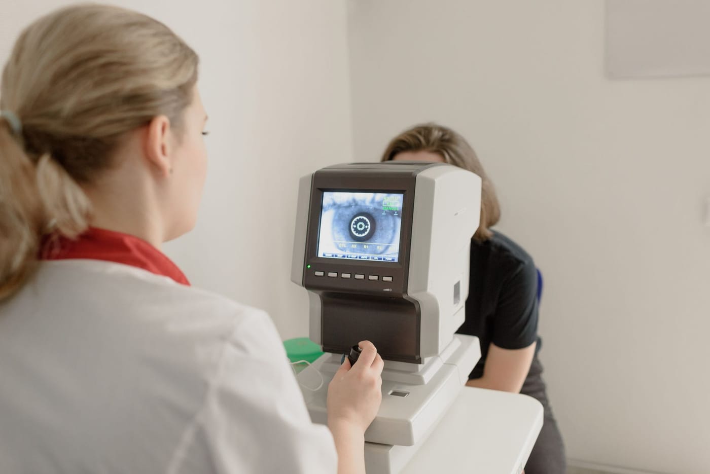
The evaluation relies on a dilated retinal exam, where a widened pupil lets the doctor see the crinkled, glistening tissue over the macula and the fine folds it creates in the retina. Optical coherence tomography, or OCT, is the key confirming test: a fast, painless cross-sectional scan that reveals the membrane, the puckering it causes, and any swelling. An Amsler grid helps map areas of distortion, and repeat OCT scans are used to judge whether the pucker is stable or advancing.
Treatment Options
If vision is good and symptoms are minor, the correct approach is observation with periodic exams and OCT imaging. No eye drop or oral medication dissolves the tissue, so careful monitoring is the standard for mild cases.
When distortion or central blur becomes bothersome, the definitive treatment is a vitrectomy with membrane peel performed by a retina surgeon. The gel inside the eye is removed and the puckering membrane is peeled off the macula, letting the retina relax and flatten. Vision and distortion usually improve gradually over weeks to months, though the result may fall short of perfect, particularly for a long-standing pucker. It is an outpatient operation, and eyes that still have their natural lens often develop a cataract afterward.
What You Should Not Do
- Do not assume every wavy-line change is a harmless pucker; distortion can also come from macular degeneration or macular swelling and deserves a look.
- Do not dismiss a sudden shower of floaters, new flashes, or a shadow in your side vision, which can mean a retinal tear or detachment.
- Do not push for surgery on a mild, stable pucker that is not affecting your daily vision.
- Do not expect stronger glasses to correct the distortion, because the issue is retinal, not refractive.
When to Call May Eye Care Center
Schedule an exam if straight lines look wavy, print seems smeared, or one eye has gradually lost sharpness, so the macula can be examined and imaged. Patients in the Hanover area rely on May Eye Care Center for this kind of assessment and follow-up. Get urgent or emergency care instead for sudden vision loss, a new curtain or shadow, or a sudden burst of floaters and flashes.
Bottom Line
A macular pucker is scar tissue that wrinkles the macula and distorts central vision; it is often mild and monitored, and when it interferes with daily sight, a membrane-peel surgery can improve it.
Frequently asked questions
01How does OCT help diagnose retina problems?
OCT imaging and a dilated retinal exam are often the key tests for retinal and macular disease. OCT can document microscopic changes in the retina that are not visible to you, and it can also be used to monitor a condition over time. Not every patient needs every test, but this kind of imaging helps determine whether a symptom is coming from the retina or from another part of the eye.
This page also answers
- What retinal symptoms are urgent?
- Can retina disease cause distortion or blind spots?
- What is the difference between macular and retinal disease?
- How does OCT help diagnose retina problems?
- When do patients need retina injections?
- When should this be checked urgently?
- What testing helps confirm the diagnosis?
- What treatments are available?
- What should patients avoid doing at home?
Medical sources
- nei.nih.gov/eye-health-information/eye-conditions-and-diseases/macular-pucker
- aao.org/eye-health/diseases/what-is-macular-pucker
This article is for educational purposes only and is not a diagnosis, treatment plan, or substitute for an eye examination by a qualified eye doctor. Eye symptoms can have many causes, and some problems can threaten vision if they are not treated promptly. Do not diagnose or treat yourself based only on online information. If you have eye pain, sudden vision loss, flashes, new floaters, a curtain or shadow in your vision, double vision, chemical exposure, trauma, severe redness, light sensitivity, or any concerning eye symptom, seek urgent medical eye care or emergency care.
Schedule your eye exam at May Eye Care Center in Hanover, PA
Serving York, Gettysburg, Adams County, and northern Maryland (driving distances). Call (717) 637-1919 or explore more about retina & vitreous at our practice.
Call (717) 637-1919