What Is a Retinal Vein Occlusion?
A retinal vein occlusion is a blockage of one of the veins that drains blood out of the retina. When the vein backs up, blood and fluid leak into the retina, causing bleeding, swelling, and usually a sudden, painless drop or blurring of vision in one eye. Treatment often includes anti-VEGF injections to reduce the swelling, along with managing the blood pressure and vascular health that contributed to the blockage.
Key Takeaways
- A retinal vein occlusion is a blocked drainage vein in the retina, which causes blood and fluid to back up into the tissue.
- It typically causes sudden, painless blurring or loss of vision in one eye, ranging from mild to severe.
- There are two main types: central retinal vein occlusion, affecting the whole retina, and branch vein occlusion, affecting one sector.
- Vision often drops because fluid collects in the macula (macular edema), which anti-VEGF injections can treat effectively.
- High blood pressure, diabetes, high cholesterol, glaucoma, and increasing age are the main risk factors, so it is partly a warning about overall vascular health.
- Sudden painless vision loss in one eye needs prompt evaluation, both to protect the eye and to check for treatable systemic risks.
Why Patients Ask This Question
Patients usually come in because vision in one eye blurred or dimmed suddenly, often noticed on waking, with no pain and no warning. Some describe it as a smudge over the center of their sight or a portion of their field that has gone hazy. Because it happens abruptly and painlessly, many are alarmed and want to know what caused it and whether the vision can come back.
What This Means for Your Eyes
The retina has arteries that bring blood in and veins that carry it out. In a vein occlusion, one of those draining veins becomes blocked, most often by a clot or by an adjacent stiffened artery pressing on it. With the exit route jammed, pressure builds in the small vessels, and blood and fluid seep into the retina, producing the scattered hemorrhages and swelling seen on exam.
The biggest threat to vision is macular edema, fluid pooling in the central macula, which blurs the sharp vision you use to read and drive. In more severe blockages, the retina can also become starved of oxygen, which can later trigger abnormal new blood vessels that cause further bleeding or a difficult form of glaucoma. How much vision is affected depends on whether the whole central vein or just a branch is involved and how much the macula swells.
Detailed Explanation
Retinal vein occlusions are divided by location. A central retinal vein occlusion blocks the main vein where it leaves the eye, affecting the entire retina and often causing more significant vision loss. A branch retinal vein occlusion blocks a smaller tributary, affecting only the sector it drains, and tends to be less severe. Doctors also classify occlusions as non-ischemic, with better blood flow and prognosis, or ischemic, with poor blood flow and a higher risk of complications.
The underlying problem is usually vascular. Chronic high blood pressure, diabetes, high cholesterol, and atherosclerosis stiffen the arteries, and where an artery crosses a vein it can compress it and promote a clot. Glaucoma and higher eye pressure raise the risk too, and in younger patients without these factors, doctors look for clotting or inflammatory conditions. Risk climbs steadily with age.
The course varies widely. Some eyes, especially with branch occlusions and mild macular edema, recover useful vision. Others have persistent swelling needing ongoing treatment, and ischemic occlusions carry the added risk of neovascularization, abnormal new vessels that can bleed or cause neovascular glaucoma months later. This is why follow-up over time matters as much as the initial treatment.
When This May Be Serious
A retinal vein occlusion is always worth prompt attention because it reflects both an eye at risk and possible uncontrolled vascular disease. Sudden, painless vision loss or blurring in one eye should be evaluated quickly rather than watched.
Specific warning signs of complications include worsening vision after an initial period of stability, new eye pain or redness, and a sudden increase in floaters, which can indicate bleeding from new abnormal vessels or a rise in eye pressure. Because vein occlusion can accompany the same conditions that cause strokes and heart attacks, any accompanying weakness, trouble speaking, or facial droop is a medical emergency requiring 911.
How an Ophthalmologist Evaluates This
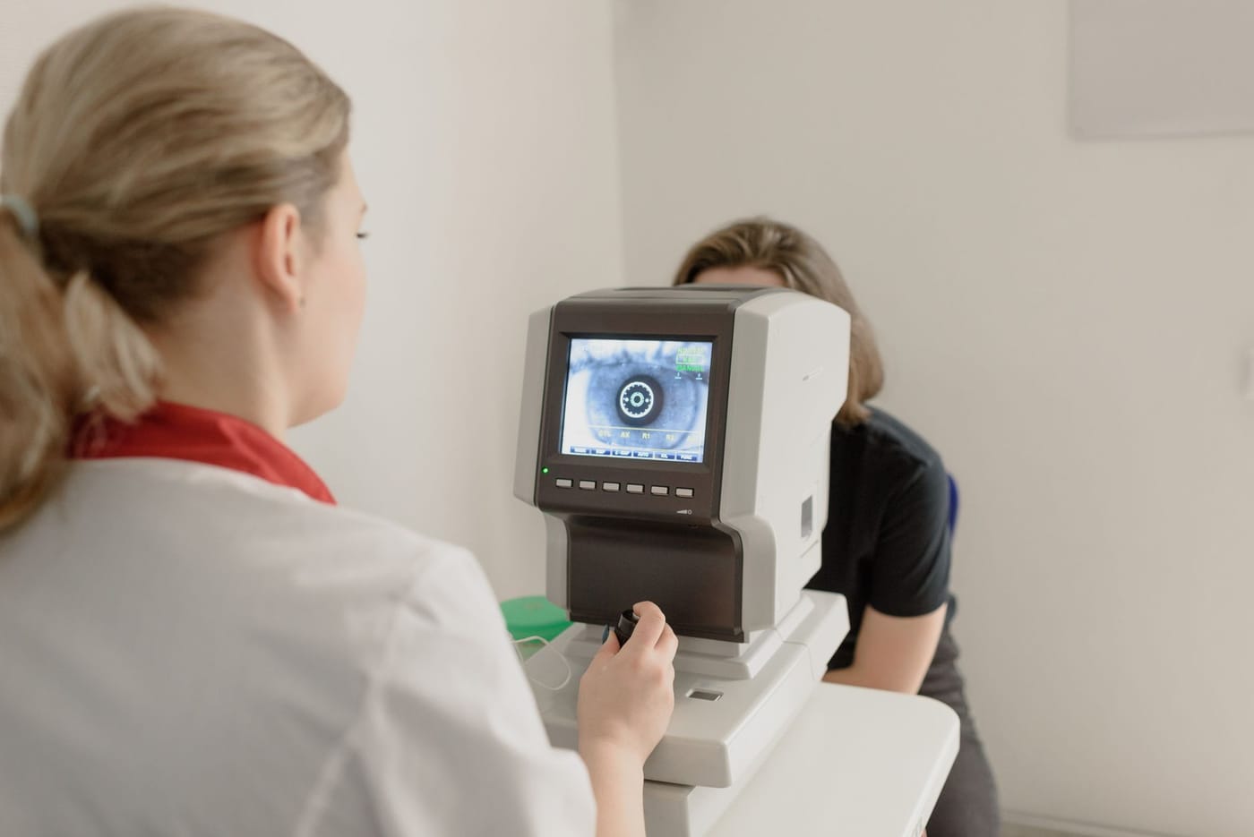
The diagnosis is made on a dilated retinal exam, where the doctor sees the characteristic pattern of scattered retinal hemorrhages, dilated tortuous veins, and swelling following the affected vein's territory. Optical coherence tomography, or OCT, measures macular edema precisely and is used to guide and monitor treatment. Fluorescein angiography, a dye-based imaging test, can map areas of poor blood flow and distinguish ischemic from non-ischemic occlusions. The workup also includes checking eye pressure and, importantly, evaluating systemic health, blood pressure, blood sugar, cholesterol, and sometimes clotting studies in younger patients, often in partnership with the primary care physician.
Treatment Options
The mainstay of treatment for vision loss from macular edema is intravitreal anti-VEGF injection, medicine given into the eye that reduces leakage and swelling and can improve vision. These are typically started on a regular schedule and then adjusted based on OCT findings. Steroid injections or implants inside the eye are another option for macular edema in selected cases.
When an occlusion is ischemic and abnormal new blood vessels develop, laser treatment (panretinal or sector photocoagulation) is used to reduce the drive for those vessels and prevent bleeding and neovascular glaucoma. Equally important is treating the underlying cause: controlling blood pressure, blood sugar, and cholesterol, and managing any glaucoma. Care is usually shared with a retina specialist for injections and with the primary physician for systemic risk factors.
What You Should Not Do
- Do not dismiss sudden painless blurring in one eye as tiredness or a passing spell; it needs prompt evaluation.
- Do not neglect blood pressure, blood sugar, or cholesterol control, since these drive the disease and threaten the other eye and your overall health.
- Do not skip follow-up visits and injections once treatment starts, because macular edema often returns if therapy stops too soon.
- Do not ignore new pain, redness, or a sudden increase in floaters weeks or months later, which can signal a serious complication.
- Do not assume glasses will help, since the blur comes from retinal swelling, not your prescription.
When to Call May Eye Care Center
Call promptly if vision in one eye blurs or dims suddenly and painlessly, so the retina can be examined, imaged with OCT, and referred for injections if needed, and so your vascular risk factors can be addressed. Patients in the Hanover area are seen at May Eye Care Center for this evaluation. If vision loss comes with weakness, slurred speech, or facial droop, treat it as a stroke and call 911 immediately.
Bottom Line
A retinal vein occlusion is a blocked drainage vein that causes sudden painless vision loss from retinal bleeding and swelling; it is commonly treated with anti-VEGF injections and by controlling the blood pressure, sugar, and cholesterol that caused it.
This page also answers
- What retinal symptoms are urgent?
- Can retina disease cause distortion or blind spots?
- What is the difference between macular and retinal disease?
- How does OCT help diagnose retina problems?
- When do patients need retina injections?
- When should this be checked urgently?
- What testing helps confirm the diagnosis?
- What treatments are available?
- What should patients avoid doing at home?
Medical sources
- aao.org/eye-health/diseases/what-is-central-retinal-vein-occlusion
- aao.org/eye-health/diseases/what-is-branch-retinal-vein-occlusion
This article is for educational purposes only and is not a diagnosis, treatment plan, or substitute for an eye examination by a qualified eye doctor. Eye symptoms can have many causes, and some problems can threaten vision if they are not treated promptly. Do not diagnose or treat yourself based only on online information. If you have eye pain, sudden vision loss, flashes, new floaters, a curtain or shadow in your vision, double vision, chemical exposure, trauma, severe redness, light sensitivity, or any concerning eye symptom, seek urgent medical eye care or emergency care.
Schedule your eye exam at May Eye Care Center in Hanover, PA
Serving York, Gettysburg, Adams County, and northern Maryland (driving distances). Call (717) 637-1919 or explore more about retina & vitreous at our practice.
Call (717) 637-1919