What Is Hypertensive Retinopathy?
Hypertensive retinopathy is damage to the small blood vessels of the retina caused by high blood pressure. It usually produces no symptoms and is found during a dilated eye exam, where the doctor sees narrowed arteries, crossing changes, and sometimes small hemorrhages or leakage. It is important because the retina offers a direct window on how high blood pressure is affecting blood vessels throughout your body, and the main treatment is getting blood pressure under good control.
Key Takeaways
- Hypertensive retinopathy is high-blood-pressure damage to the retina's small blood vessels.
- It is often silent, causing no symptoms, and is frequently discovered on a routine dilated eye exam.
- Early signs include narrowed retinal arteries and changes where arteries cross veins; more advanced signs include hemorrhages, cotton-wool spots, and swelling.
- It is a marker of systemic vascular risk, reflecting the same stress on blood vessels in the heart, brain, and kidneys.
- The core treatment is controlling blood pressure, not an eye-specific procedure.
- Severe, sudden findings such as optic nerve swelling can indicate dangerously high blood pressure (a hypertensive emergency) that needs immediate care.
Why Patients Ask This Question
Most people ask because an eye doctor mentioned signs of high blood pressure in the back of their eye, sometimes before their primary doctor had flagged a blood pressure problem, and they are surprised the eye can reveal that. Others have blurred vision or a known history of hypertension and want to know whether it is harming their sight. Because it is usually painless and silent, the news often comes as a genuine surprise.
What This Means for Your Eyes
The retina is unique because it is the one place a doctor can directly see small blood vessels without surgery. When blood pressure runs high over time, the tiny retinal arteries thicken and narrow to protect themselves, and where they cross veins they can pinch them. With more severe or rapidly rising pressure, vessels can leak, producing small hemorrhages, fluffy white cotton-wool spots from blocked blood flow, and deposits of leaked material.
In most people, mild hypertensive retinopathy does not by itself blur vision. The greater significance is what it implies about the rest of the body, because the same vascular stress is happening in the heart, brain, and kidneys. Vision can be affected when high blood pressure causes swelling of the macula, bleeding, or optic nerve swelling, or when it contributes to a retinal vein or artery occlusion.
Detailed Explanation
Hypertensive retinopathy develops as chronically elevated blood pressure forces the retinal arteries to constrict and their walls to thicken. Doctors describe a spectrum of changes: early on, generalized and focal arterial narrowing and arteriovenous crossing changes (where a stiffened artery indents the vein it crosses). As disease advances, the vessels can leak, causing flame-shaped hemorrhages, cotton-wool spots (small areas of nerve-fiber layer damage from poor perfusion), and hard exudates. In the most severe form, malignant or accelerated hypertension, the optic nerve head swells, which signals a hypertensive emergency.
The key risk factor is the height and duration of blood pressure elevation, amplified by diabetes, high cholesterol, smoking, and age. High blood pressure also predisposes to related retinal problems, retinal vein occlusion, retinal artery occlusion, and worsening of diabetic retinopathy, so it rarely acts alone.
The typical course tracks blood pressure control. When pressure is brought down and kept in a healthy range, mild retinal changes often stabilize or partially improve, and the risk of complications falls. Poorly controlled hypertension, by contrast, allows the vascular damage to progress in the eye and elsewhere in the body.
When This May Be Serious
Mild hypertensive retinopathy is usually a warning sign rather than an immediate threat to vision, but certain findings are serious. Swelling of the optic nerve, extensive hemorrhages, or a sudden drop in vision can indicate severely elevated blood pressure (a hypertensive emergency) that needs urgent medical care, sometimes in the emergency room.
Seek prompt eye evaluation for sudden painless vision loss, new distortion, or a shadow in your vision, which can mean high blood pressure has caused a vein or artery occlusion or macular swelling. And any accompanying signs of stroke, weakness, facial droop, or trouble speaking, are a 911 emergency.
How an Ophthalmologist Evaluates This
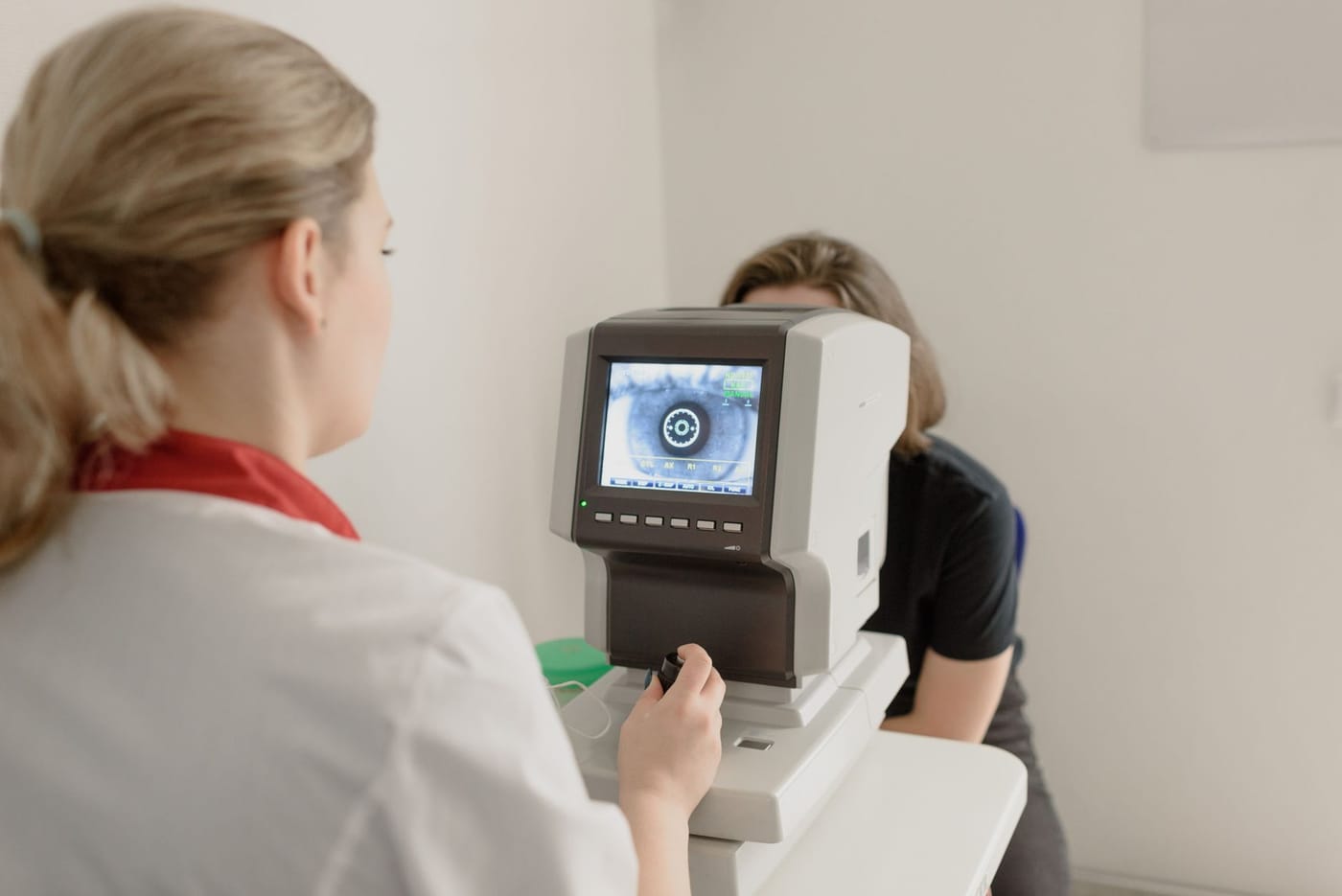
The diagnosis is made on a dilated retinal exam, where the doctor grades the retinal arteries for narrowing, examines the points where arteries cross veins, and looks for hemorrhages, cotton-wool spots, exudates, and any swelling of the optic nerve or macula. Blood pressure is measured, and findings are correlated with your medical history. Optical coherence tomography, or OCT, can document macular swelling if vision is affected, and photographs are useful for tracking changes over time. Importantly, the eye doctor communicates with your primary care physician, because managing the blood pressure itself is central to treatment.
Treatment Options
The primary treatment for hypertensive retinopathy is not done in the eye at all, it is achieving and maintaining good blood pressure control through medication, diet, exercise, weight management, and reducing salt, in partnership with your primary care doctor. As blood pressure improves, mild retinal changes often stabilize or partly reverse, and the risk of serious complications drops.
Eye-directed treatment is reserved for complications. If high blood pressure leads to macular swelling or a retinal vein occlusion, anti-VEGF injections may be used; if it contributes to diabetic retinopathy, laser or injections may be needed. Severely elevated blood pressure with optic nerve swelling is a medical emergency requiring urgent blood pressure lowering under medical supervision. Regular eye exams help monitor the retina, especially in people with diabetes or long-standing hypertension.
What You Should Not Do
- Do not assume normal vision means your blood pressure is fine, hypertensive retinopathy is usually silent and can be present without symptoms.
- Do not stop or skip blood pressure medications, since consistent control is the actual treatment for this condition.
- Do not ignore sudden vision loss, new distortion, or a shadow, which can signal a vein or artery occlusion from high blood pressure.
- Do not treat a severe headache with visual changes and very high blood pressure as minor, it can be a hypertensive emergency.
- Do not rely on eye drops or glasses to fix this, the fix is systemic blood pressure control.
When to Call May Eye Care Center
Schedule a dilated exam if you have high blood pressure, or if a doctor has mentioned vascular changes in your eyes, so the retina can be checked and monitored; patients in the Hanover area are seen at May Eye Care Center for this. Seek urgent care for sudden vision loss, new distortion, or a shadow in your vision, and call 911 if very high blood pressure comes with severe headache, chest pain, or stroke symptoms.
Bottom Line
Hypertensive retinopathy is high-blood-pressure damage to the retina's vessels that is usually silent but signals body-wide vascular risk; the main treatment is good blood pressure control, with eye-specific care reserved for complications.
This page also answers
- What retinal symptoms are urgent?
- Can retina disease cause distortion or blind spots?
- What is the difference between macular and retinal disease?
- How does OCT help diagnose retina problems?
- When do patients need retina injections?
- When should this be checked urgently?
- What testing helps confirm the diagnosis?
- What treatments are available?
- What should patients avoid doing at home?
Medical sources
This article is for educational purposes only and is not a diagnosis, treatment plan, or substitute for an eye examination by a qualified eye doctor. Eye symptoms can have many causes, and some problems can threaten vision if they are not treated promptly. Do not diagnose or treat yourself based only on online information. If you have eye pain, sudden vision loss, flashes, new floaters, a curtain or shadow in your vision, double vision, chemical exposure, trauma, severe redness, light sensitivity, or any concerning eye symptom, seek urgent medical eye care or emergency care.
Schedule your eye exam at May Eye Care Center in Hanover, PA
Serving York, Gettysburg, Adams County, and northern Maryland (driving distances). Call (717) 637-1919 or explore more about retina & vitreous at our practice.
Call (717) 637-1919