What Is Corneal Topography?
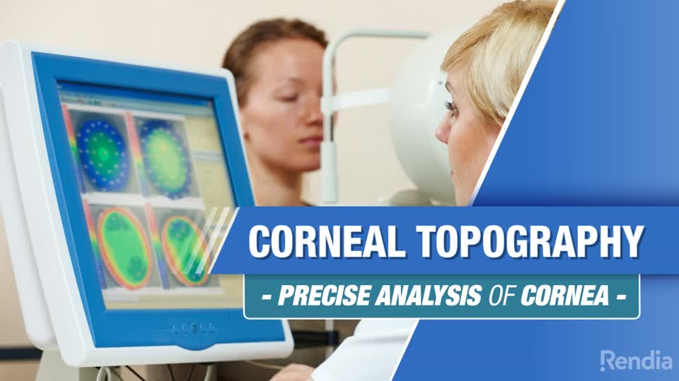
Corneal topography is a quick, non-contact test that creates a detailed three-dimensional map of the curvature of your cornea, the clear front window of the eye. Rings of light are projected onto the corneal surface, and a computer measures how they reflect to chart every steep and flat area, much like a topographic map shows the hills and valleys of a landscape. There are no drops, no puff of air, and nothing touches your eye. The map is used to fit specialty contact lenses, screen and plan refractive surgery such as LASIK, and diagnose conditions that distort the corneal shape, like astigmatism and keratoconus.
§Read video transcript
Corneal topography is the latest technology to ensure precise analysis of your cornea, it is used for advanced contact lens fittings, refractive surgery, and for diagnosing, monitoring, and treating various eye conditions. The cornea is the clear outer surface "window" of the eye. It transmits and focuses light into the eye and plays a major role in how you see. Corneal topography creates a 3D map of the curvature of the cornea. Just as the Earth appears to be a perfectly smooth sphere from outer space, the cornea looks like a perfect curve with the naked eye, however, a closer inspection reveals that some parts are steep and others flat. Using corneal topography, your doctor can closely inspect and map the valleys and hills that characterize your cornea’s shape. This sort of 3D map allows for the detection of conditions that would be invisible with conventional testing. During the test, rings of light are projected onto the cornea, and measurements are taken of your eye’s surface. This test is non-invasive. There are no drops, there is no contact with the eye, and no puff of air to worry about. Your doctor will use the computer-generated map to create a custom, precise treatment for your eye health and vision needs. For example - if a contact lens is too tight, it could constrict normal tear flow - or if too loose, it could cause excess movement and damage the corneal tissue. Corneal topography helps your eye care professional identify your unique corneal shape, and find the best solution for you.
Key Takeaways
- Corneal topography maps the exact shape and curvature of the cornea in 3D, revealing detail invisible to the naked eye.
- It is completely non-invasive: rings of light are reflected off the eye, with no drops, no contact, and no air puff.
- It is essential for advanced contact lens fittings, for screening and planning LASIK, and for diagnosing corneal shape disorders.
- It detects and tracks keratoconus, a progressive thinning and bulging of the cornea, often before it noticeably affects vision.
- It explains irregular or hard-to-correct astigmatism that ordinary glasses cannot fully sharpen.
- A distorted or rapidly changing map can warn that a cornea is not a safe candidate for laser vision surgery.
Why Patients Ask This Question
Patients usually meet this test when exploring LASIK, being fitted for hard-to-fit contact lenses, or being evaluated because their vision cannot be fully sharpened with standard glasses. It looks unusual, with rings of light and a color map, so people want to know what it measures and whether it will be uncomfortable. Some are anxious after hearing the word keratoconus and want to understand what the mapping can tell them.
What This Means for Your Eyes
The cornea is the eye's clear outer surface, and it does most of the focusing of light before it reaches the lens. To the naked eye it looks like a smooth, even dome, but under topography its true shape emerges, with subtle steep and flat regions that determine how well light comes to a focus. Even small irregularities can blur vision or make it hard to correct.
Because the cornea's curvature is so central to clear sight, mapping it precisely tells your doctor a great deal. An evenly curved cornea focuses light cleanly, while an irregular or cone-shaped cornea scatters it, causing astigmatism, distortion, or glare. Detecting those irregularities is what allows the right lens, or the right surgical decision, to be matched to your unique eye.
Detailed Explanation
During the test, a lighted pattern of concentric rings is projected onto the cornea, and a camera captures how they reflect off its surface. A computer analyzes the reflection point by point and produces a color-coded map, where warmer colors typically mark steeper areas and cooler colors flatter ones. Because it reads the shape optically, the test is fast and requires nothing more than looking at a target.
The map detects conditions conventional testing can miss. Keratoconus, in which the cornea progressively thins and bulges into a cone, produces a characteristic steepening that topography can catch early and then monitor. Irregular astigmatism, corneal scarring, and shape changes from prior surgery or long-term contact lens wear also show up clearly. In contact lens fitting, the map guides the right curvature: a lens too tight can restrict tear flow, while one too loose moves excessively and can irritate the corneal surface. In refractive surgery, it helps confirm a cornea is regular and thick enough to treat safely and guides a precise, custom plan for each eye.
When This May Be Serious
The scan is entirely benign, and corneal shape disorders are usually not emergencies, but some findings matter. A pattern suggesting keratoconus, or a cornea too thin or irregular for laser surgery, changes management and may steer you away from LASIK toward safer options. Rapid change over time can signal progressing keratoconus that may benefit from treatment to halt it. Separately, and unrelated to topography, sudden severe eye pain, a rapid drop in vision, or a corneal injury with light sensitivity and redness are urgent problems that need prompt care.
How an Ophthalmologist Evaluates This
Corneal topography is interpreted alongside the rest of the corneal and refractive evaluation. Your ophthalmologist compares the map with your refraction and best-corrected vision, examines the cornea at the slit lamp, and, when screening for surgery or keratoconus, often adds a measurement of corneal thickness. For suspected keratoconus, the shape map is combined with those thickness readings and followed over time to judge whether the cornea is stable or progressing. For a contact lens fitting, the map defines the curvature the lens must match.
Treatment Options
Topography is a mapping test, so it guides treatment rather than being one. A regular cornea may simply confirm you are a good candidate for glasses, contacts, or laser correction and help design a precise plan. Irregular astigmatism may be corrected with specialty contact lenses such as rigid gas-permeable or scleral lenses that glasses cannot match. Early or progressing keratoconus can be treated with corneal cross-linking to strengthen the cornea and slow the bulging, and specialty lenses or, in advanced cases, a corneal transplant may be considered. In each situation, the map matches the right option to your cornea's shape.
What You Should Not Do
- Do not pursue LASIK without corneal mapping; skipping it removes a key safeguard against operating on a thin or irregular cornea.
- Do not ignore worsening or hard-to-correct vision as just a stronger prescription; it can be early keratoconus that a map would catch.
- Do not rub your eyes vigorously if you have keratoconus, as habitual hard rubbing is linked to its progression.
- Do not assume a specialty contact lens fit can be judged by trial and error alone; the map makes the fit safer and more accurate.
When to Call May Eye Care Center
Consider a visit if you are exploring LASIK, if your vision cannot be fully corrected with glasses, if you have been told you might have keratoconus, or if you need a specialty contact lens fitting. Sudden severe pain, a sharp drop in vision, or a corneal injury should be seen right away. May Eye Care Center offers corneal topography on site for patients in the Hanover, Pennsylvania area.
Bottom Line
Corneal topography is a painless, no-contact scan that maps the true shape of your cornea, letting your doctor fit difficult contact lenses, screen and plan laser vision correction safely, and catch shape disorders like keratoconus early.
This page also answers
- What eye tests detect glaucoma or retina disease?
- Why did my eye doctor order an OCT or visual field?
- Do eye tests hurt?
- How often should eye tests be repeated?
- What do eye imaging results mean?
- When should this be checked urgently?
- What testing helps confirm the diagnosis?
- What treatments are available?
- What should patients avoid doing at home?
Medical sources
- aao.org/eye-health/tips-prevention/eye-exams-101
- aao.org/eye-health/a-z
- nei.nih.gov/eye-health-information/healthy-vision
This article is for educational purposes only and is not a diagnosis, treatment plan, or substitute for an eye examination by a qualified eye doctor. Eye symptoms can have many causes, and some problems can threaten vision if they are not treated promptly. Do not diagnose or treat yourself based only on online information. If you have eye pain, sudden vision loss, flashes, new floaters, a curtain or shadow in your vision, double vision, chemical exposure, trauma, severe redness, light sensitivity, or any concerning eye symptom, seek urgent medical eye care or emergency care.
Schedule your eye exam at May Eye Care Center in Hanover, PA
Serving York, Gettysburg, Adams County, and northern Maryland (driving distances). Call (717) 637-1919 or explore more about LASIK at our practice.
Call (717) 637-1919