What Is Keratoconus?
Keratoconus is a progressive condition in which the cornea, the clear dome at the front of the eye, gradually thins and bulges outward into a cone shape. That irregular shape distorts light and causes blurred, often worsening vision, along with light sensitivity, glare, and trouble seeing at night. It usually begins in the teens to mid-twenties, tends to run in families, and can range from mild to severe. Modern treatment can halt its progression when it is caught early, which is why timely diagnosis matters.
§Read video transcript
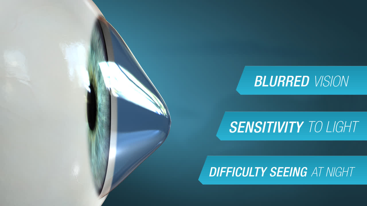
Keratoconus is the thinning of the cornea the clear dome-shaped outer surface of your eye. Eventually, the cornea bulges outward into a cone shape causing blurred vision, sensitivity to light and difficulty seeing at night. Most commonly affecting individuals between the ages of 10 to 25, the chance of developing it increases if it runs in the family. Different treatment options are available, depending on the severity. Mild to Moderate Keratoconus may be treated with eyeglasses or contact lenses. Another treatment is called Corneal Collagen Cross-linking, which uses riboflavin and UV light to strengthen the cornea. Often this treatment is used in addition to the insertion of Intacs. Intacs corneal implants are two small crescent shaped pieces made of plastic polymer which are inserted into the cornea to support the cornea’s shape, leading to improved vision. But if the cornea becomes scarred, making it painful to wear contacts, surgery is another option. Recovery time can take up to one year and you may need to continue wearing rigid contacts to ensure clear vision. Keratoconus can worsen over time so early detection is key. If you notice any of the symptoms, it is best to schedule an appointment with your eye care professional. He or she can work with you, to make sure you have the best vision possible!
Key Takeaways
- The cornea thins and steepens into a cone, scattering light and blurring vision.
- It most often starts between about ages 10 and 25 and is more likely if it runs in the family.
- Early symptoms include blurred and distorted vision, glare, halos, light sensitivity, and frequent changes in the eyeglass prescription.
- Vigorous eye rubbing is linked to progression and should be avoided.
- It can worsen over time, so early detection is key; corneal cross-linking can halt progression before vision is badly affected.
Why Patients Ask This Question
Many patients are young and frustrated that their glasses never seem to work well: their prescription keeps changing, vision is blurry or doubled even with correction, and lights streak and glare, especially at night. Some are told during an eye exam that their cornea has an unusual shape. They want to understand what keratoconus is, why it is happening to them, and whether their sight can be protected.
What This Means for Your Eyes
The cornea does most of the eye's focusing, and it works best when it is a smooth, regular dome. In keratoconus the cornea weakens and thins, so the normal eye pressure pushes it outward into a cone. That cone-shaped surface bends light unevenly, producing irregular astigmatism, which glasses cannot fully correct because they only fix regular shapes.
As a result, vision becomes blurred and distorted, straight lines may look wavy, and lights develop halos and streaks that make night driving hard. In the early stages, stronger or frequently changing glasses may keep up, but as the cone steepens, glasses stop working and rigid or specialty contact lenses are needed to create a smooth optical surface. In advanced cases the cornea can scar, which further blurs vision and can make contact lenses uncomfortable to wear.
Detailed Explanation
Keratoconus develops when the structural fibers of the cornea weaken, allowing the front of the eye to thin and bulge. It usually appears in adolescence or early adulthood and often progresses over years before stabilizing, though the rate varies widely from person to person. A family history raises the risk, and it is associated with chronic eye rubbing, allergic eye disease, and certain conditions such as Down syndrome and connective tissue disorders. Vigorous eye rubbing, in particular, is thought to mechanically weaken the cornea and speed progression.
The typical course begins with increasing nearsightedness and astigmatism that prompts frequent prescription changes. As the cone steepens, glasses no longer give clear vision and specialty contact lenses become the mainstay of correction. Progression tends to be faster in younger patients. In a minority of cases the cornea scars or, rarely, swells suddenly (a condition called hydrops), causing an abrupt drop in vision. Because damage from steepening and scarring is not reversible, the goal of modern care is to detect the condition early and stop it from advancing.
When This May Be Serious
Keratoconus is not an emergency in the usual sense, but certain changes deserve prompt attention:
- A sudden, marked drop in vision with eye pain, redness, and light sensitivity can signal corneal hydrops (sudden corneal swelling) and should be evaluated quickly.
- Rapidly worsening vision or frequently changing prescriptions, especially in a young patient, suggests active progression that may benefit from cross-linking.
- Contact lenses that become painful or impossible to wear may mean the cornea is scarring.
Most importantly, progressive keratoconus that is not treated can lead to permanent vision loss, so worsening vision should not simply be watched.
How an Ophthalmologist Evaluates This
The key test is corneal topography (and tomography), which maps the shape and steepness of the cornea and can reveal keratoconus even in early stages before it is obvious. These maps also measure corneal thickness and detect thinning. At the slit lamp, the doctor looks for the classic signs of a steepened, thinned cornea and any scarring, and measures how much vision can be corrected with glasses versus specialty lenses. Repeat topography over time shows whether the condition is stable or progressing, which directly guides whether cross-linking is recommended. Because it often affects both eyes to different degrees, both are examined and monitored.
Treatment Options
Treatment is matched to severity. Mild to moderate keratoconus can often be corrected with eyeglasses or contact lenses. As the cornea steepens, rigid gas-permeable or scleral contact lenses create a smooth front surface and restore clearer vision when glasses no longer can.
To stop progression, the cornea can be strengthened with corneal collagen cross-linking, which uses riboflavin (vitamin B2) drops activated by ultraviolet light to reinforce the bonds within the cornea. Cross-linking does not reverse the cone, but it halts further steepening in most patients, so it is most valuable when the disease is caught early. Intacs, small crescent-shaped corneal implants, can be inserted to support the cornea's shape and improve vision, and are sometimes combined with cross-linking. If the cornea becomes badly scarred or too irregular for comfortable lens wear, a corneal transplant is an option; recovery can take up to a year, and rigid contact lenses may still be needed afterward for the sharpest vision.
What You Should Not Do
- Do not rub your eyes; vigorous rubbing is linked to worsening keratoconus.
- Do not ignore steadily worsening vision or a rapidly changing prescription, since progression that is caught early can be halted.
- Do not assume glasses alone are the answer once vision is distorted; specialty contact lenses often do far better.
- Do not leave allergies or itchy eyes untreated, because the urge to rub can drive the disease.
- Do not skip follow-up visits, as monitoring the corneal shape is how progression is detected in time to treat it.
When to Call May Eye Care Center
Call for an evaluation if you are a young adult with blurry, distorted vision that glasses do not fully correct, or if your astigmatism keeps changing, so the corneal shape can be mapped. Patients in the Hanover area can be assessed and, if appropriate, offered cross-linking to protect their vision. Seek prompt care for a sudden drop in vision with a painful, red, light-sensitive eye.
Bottom Line
Keratoconus is a progressive thinning and coning of the cornea that distorts vision and usually starts in the teens or twenties, but early detection and cross-linking can halt it before it damages sight. If your vision is blurry and glasses are not enough, have your corneas evaluated at May Eye Care Center.
Frequently asked questions
01Can a corneal problem blur vision?
Yes, corneal disease is one of the conditions that can blur vision, including scratches, infection, scarring, and problems with corneal shape. Many other issues, from dry eye to early cataract, glaucoma, or retina disease, can blur vision as well, which is why a new change in vision deserves a real eye examination rather than self-diagnosis.
This page also answers
- Why does my eye feel scratched or irritated?
- Can a corneal problem blur vision?
- When is a painful red eye urgent?
- How does an eye doctor examine the cornea?
- Can corneal disease require surgery?
- When should this be checked urgently?
- What testing helps confirm the diagnosis?
- What treatments are available?
- What should patients avoid doing at home?
Medical sources
This article is for educational purposes only and is not a diagnosis, treatment plan, or substitute for an eye examination by a qualified eye doctor. Eye symptoms can have many causes, and some problems can threaten vision if they are not treated promptly. Do not diagnose or treat yourself based only on online information. If you have eye pain, sudden vision loss, flashes, new floaters, a curtain or shadow in your vision, double vision, chemical exposure, trauma, severe redness, light sensitivity, or any concerning eye symptom, seek urgent medical eye care or emergency care.
Schedule your eye exam at May Eye Care Center in Hanover, PA
Serving York, Gettysburg, Adams County, and northern Maryland (driving distances). Call (717) 637-1919 or explore more about cornea at our practice.
Call (717) 637-1919