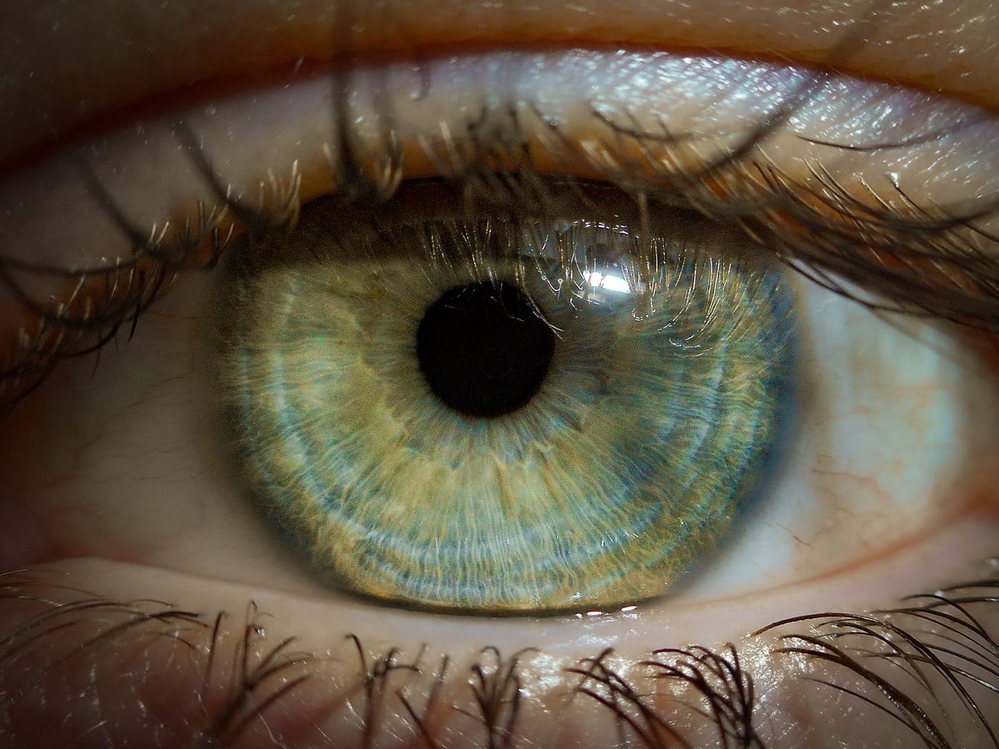
What Is Recurrent Corneal Erosion?
Recurrent corneal erosion is a condition where the outer skin of the cornea (the epithelium) repeatedly breaks loose because it never bonded firmly to the layer beneath it. It typically follows a past scratch, especially from a fingernail or paper, or occurs with a corneal dystrophy. The hallmark is sudden, sharp pain, tearing, and light sensitivity, classically on first opening the eyes in the morning, that recurs over weeks to months. It is not dangerous to sight in most cases but can be very disruptive, and it is treatable.
Key Takeaways
- The eye's surface skin repeatedly detaches because it is poorly anchored to the tissue below.
- Attacks often follow a prior corneal abrasion, particularly a fingernail or paper cut, and can begin months or years later.
- The classic symptom is sharp pain on waking and opening the eyes, with tearing, redness, and light sensitivity.
- Episodes can recur for weeks to months and range from mild irritation to a full painful abrasion.
- It is usually not sight-threatening, but recurring severe pain, blurred vision, or signs of infection warrant evaluation.
Why Patients Ask This Question
These patients describe a puzzling, repeating pattern: they open their eyes in the morning and are hit with sudden, stabbing pain, watering, and light sensitivity, sometimes for just seconds, sometimes lasting hours or days. Often it started long after a minor eye scratch they had nearly forgotten. They want to understand why the same eye keeps flaring up and whether it can be stopped.
What This Means for Your Eyes
The corneal epithelium is meant to be glued firmly to the layer underneath it by tiny anchoring structures. In recurrent erosion, that attachment is weak, so the surface skin can peel away, especially overnight. During sleep the surface dries slightly and can stick to the inside of the eyelid; when you open your eyes in the morning, the loosely attached epithelium is pulled off, exposing the nerve-rich tissue underneath and causing sudden sharp pain.
Because the same weak spot keeps giving way, the problem recurs. Between attacks the eye may feel completely normal. Vision is usually preserved, though it can blur during an episode or if the surface heals irregularly. The condition is best thought of as a mechanical anchoring problem of the surface rather than a progressive disease, which is reassuring, though the recurring pain can be genuinely disabling until it is treated.
Detailed Explanation
The most common trigger is a previous corneal abrasion, especially a sharp injury from a fingernail, paper edge, or plant, which can leave the healed epithelium loosely attached at that site. Episodes may begin weeks, months, or occasionally years after the original scratch. The other major cause is an inherited corneal condition, such as epithelial basement membrane (map-dot-fingerprint) dystrophy, in which the anchoring layer is abnormal from the start; in these patients both eyes can be affected. Dry eye worsens the problem by letting the surface stick to the lid overnight.
Attacks tend to strike on waking because the lids have been closed and the tear film thinned during sleep. A mild episode may be a fleeting stab that settles quickly; a severe one can strip a sizable patch of surface, producing a full-blown painful abrasion that takes days to heal, only to recur later. Over time and with proper treatment, the anchoring can improve, but many patients need a deliberate plan to strengthen the attachment and stop the cycle.
When This May Be Serious
Recurrent erosion is usually not a threat to sight, but seek care if:
- Pain is severe or attacks are frequent and disrupting your life.
- Vision becomes blurred and does not clear between episodes.
- The eye develops increasing redness, discharge, or a white spot, which can signal infection of the exposed surface.
- Episodes are not responding to lubrication.
An exposed corneal surface can occasionally become infected, so a red, painful eye with worsening symptoms rather than the usual brief morning flare should be checked promptly.
How an Ophthalmologist Evaluates This
Diagnosis often comes from the story alone, the classic sharp pain on opening the eyes in the morning, but it is confirmed at the slit lamp. The doctor examines the corneal surface, sometimes catching an area of loose or heaped-up epithelium, and uses fluorescein dye to reveal a current erosion or the subtle irregularities of a dystrophy. Both eyes are examined, because a basement membrane dystrophy that causes erosions in one eye is frequently present in the other. The doctor also asks about any past eye injury, since a long-ago scratch is a common clue.
Treatment Options
First-line treatment aims to keep the surface moist and encourage firmer reattachment. This means generous artificial tears during the day and, importantly, a lubricating ointment or hypertonic (salt) ointment at bedtime for weeks to months so the surface does not stick to the lid overnight. Treating any underlying dry eye helps. During an acute erosion, the eye is managed like an abrasion, sometimes with a bandage contact lens placed by the doctor for comfort while it heals.
When episodes keep recurring despite lubrication, in-office procedures strengthen the anchoring. Anterior stromal puncture places tiny spots that promote firm scarring-down of the surface, and phototherapeutic keratectomy (PTK), a laser treatment, smooths and resurfaces the cornea to help the epithelium adhere. These procedures are effective at reducing or ending recurrences for most patients.
What You Should Not Do
- Do not rub the eye, especially on waking, which can tear off the loosely attached surface.
- Do not skip your nightly lubricating ointment once it is prescribed; consistency over weeks is what breaks the cycle.
- Do not let the eyes get dry, and treat dry eye actively, since dryness triggers attacks.
- Do not open your eyes abruptly on waking; blinking gently or moistening them first can help.
- Do not ignore new redness, discharge, or worsening pain, which could mean the exposed surface has become infected.
When to Call May Eye Care Center
Call if you have repeated episodes of sharp eye pain on waking, or a scratch that keeps coming back, so the surface can be examined and a treatment plan started. Patients in the Hanover area can be evaluated and, when needed, offered in-office procedures. Seek prompt care for a severely painful red eye with discharge or a white spot, which suggests infection rather than a simple erosion.
Bottom Line
Recurrent corneal erosion is a repeating breakdown of the eye's surface skin, usually after an old scratch, that causes classic sharp pain on waking and responds well to lubrication and, if needed, minor procedures. Persistent or severe episodes should be evaluated at May Eye Care Center.
This page also answers
- Why does my eye feel scratched or irritated?
- Can a corneal problem blur vision?
- When is a painful red eye urgent?
- How does an eye doctor examine the cornea?
- Can corneal disease require surgery?
- When should this be checked urgently?
- What testing helps confirm the diagnosis?
- What treatments are available?
- What should patients avoid doing at home?
Medical sources
This article is for educational purposes only and is not a diagnosis, treatment plan, or substitute for an eye examination by a qualified eye doctor. Eye symptoms can have many causes, and some problems can threaten vision if they are not treated promptly. Do not diagnose or treat yourself based only on online information. If you have eye pain, sudden vision loss, flashes, new floaters, a curtain or shadow in your vision, double vision, chemical exposure, trauma, severe redness, light sensitivity, or any concerning eye symptom, seek urgent medical eye care or emergency care.
Schedule your eye exam at May Eye Care Center in Hanover, PA
Serving York, Gettysburg, Adams County, and northern Maryland (driving distances). Call (717) 637-1919 or explore more about cornea at our practice.
Call (717) 637-1919