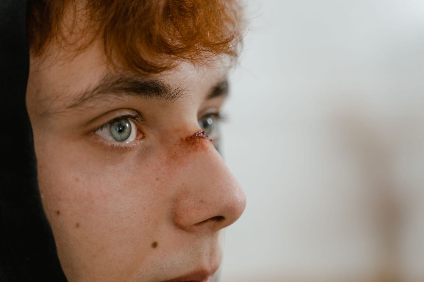
What Is a Hyphema?
A hyphema is blood pooling in the anterior chamber, the space between the clear cornea and the colored iris, usually after a blunt blow to the eye. You may see a reddish tint or an actual layer of blood settling across the bottom of the colored part of the eye, along with blurred vision, pain, and light sensitivity. It is treated as urgent because the same injury can raise the pressure inside the eye and because a rebleed in the first few days can be worse than the original. The eye needs prompt examination, rest with the head elevated, a protective shield, and close monitoring, so it should be seen the same day.
Key Takeaways
- A hyphema is blood inside the front chamber of the eye, typically from blunt trauma.
- You may see blood layered across the lower part of the iris, with blur, ache, and light sensitivity.
- The two main dangers are a spike in eye pressure and a rebleed, which often happens in the first few days.
- Management centers on rest, keeping the head elevated, a rigid shield, and avoiding straining and blood thinners.
- People with sickle cell trait or disease are at higher risk of pressure problems and need special care.
- This is urgent: a hyphema needs a same-day exam and follow-up, not watchful waiting at home.
Why Patients Ask This Question
After a ball, fist, or airbag hits the eye, patients notice the colored part looks 'filled with blood' or see a red line pooling at the bottom of the eye, and it is frightening. They want to know what that blood means, whether it will clear, and how serious it is, especially since the eye can ache and the vision can be hazy.
What This Means for Your Eyes
The anterior chamber is normally filled with clear fluid that drains out through a filter at the angle where the iris meets the cornea. In a hyphema, blunt force tears small vessels of the iris or ciliary body, and blood escapes into that fluid. Because blood is heavier than the fluid, it settles into a visible layer when you are upright, which is why sitting up with the head raised helps it collect low and clear.
The blood itself will usually reabsorb, but the risks come from what it does meanwhile. Red cells and debris can clog the drainage angle and drive up eye pressure, which if severe and prolonged can damage the optic nerve. And the injured vessel can bleed again as the clot breaks down, often around days three to five, sometimes filling more of the chamber than before. Those two risks, pressure and rebleeding, are why a hyphema is watched closely rather than left alone.
Detailed Explanation
Most hyphemas follow blunt trauma, a sports ball, a fist, an elbow, a paintball, an airbag, but they can also occur after surgery or from abnormal vessels in the eye. They are graded by how much of the anterior chamber the blood fills, from a thin layer to a chamber that is completely full (a 'total' or 'eight-ball' hyphema), with higher grades carrying more risk to pressure and vision.
The course runs over several days to two weeks as the blood clears. The critical window for a rebleed is roughly the first several days, as the initial clot retracts. Elevated eye pressure can appear early or later from blood clogging the drain. Patients with sickle cell trait or disease are especially vulnerable: their red cells sickle in the low-oxygen environment of the anterior chamber, block the drain more easily, and tolerate high pressure poorly, so even a small hyphema is managed carefully. A significant hyphema also means the eye took enough force to warrant checking for angle damage that can raise pressure years later.
When This May Be Serious
A hyphema is already urgent; seek care immediately, and treat these as red flags of a worsening problem:
- Severe or increasing eye pain, or pain with nausea and vomiting, which can signal a dangerous pressure spike.
- Vision that is getting worse, or blood that is filling more of the eye (a rebleed).
- A new headache or halos around lights with the painful eye.
- Known sickle cell trait or disease, or use of blood thinners, which raise the risk.
- Any hyphema at all, because it needs prompt examination and cannot be safely judged at home.
How an Ophthalmologist Evaluates This
The doctor measures vision and examines the eye at the slit-lamp to grade how much blood is in the chamber and to look for other trauma to the iris, lens, and cornea. Eye pressure is measured, and rechecked over the following days because pressure can climb as the eye clears. The pupil is dilated when safe to examine the retina for accompanying injury, and gonioscopy, a lens that views the drainage angle, may be done later to check for angle recession once the eye has settled. Sickle cell status is checked in at-risk patients, and CT imaging is added if the mechanism suggests a fracture or a ruptured globe.
Treatment Options
The foundation is rest with the head of the bed elevated so the blood settles and clears, along with a rigid eye shield and limited activity to reduce the chance of a rebleed. A dilating drop keeps the eye comfortable and steadies the iris, and a topical steroid calms inflammation. If eye pressure rises, pressure-lowering drops or pills are used; pressure control is especially aggressive in sickle cell patients, in whom certain drops are avoided. Blood thinners and strenuous activity are held. Most hyphemas clear with this approach, but a chamber that stays full, an uncontrolled pressure, or blood staining the cornea may require surgery to wash out the blood.
What You Should Not Do
- Do not carry on normal activity; genuine rest with the head elevated is part of the treatment.
- Do not lift, bend, strain, or exercise, which can trigger a rebleed.
- Do not take aspirin or other blood-thinning pain relievers unless your doctor specifically approves them.
- Do not rub, press on, or remove the eye shield except as instructed.
- Do not skip the follow-up visits; the pressure checks and rebleed watch over the next several days are the whole point.
When to Call May Eye Care Center
A hyphema needs to be seen the same day. Call May Eye Care Center right away after any blunt eye injury that leaves blood in the eye, or go to an emergency room if severe pain, vomiting, or rapidly worsening vision suggests a pressure spike. Patients in the Hanover area can reach the office to arrange the prompt exam and the close follow-up a hyphema requires over the first days.
Bottom Line
A hyphema is blood in the front chamber of the eye after blunt trauma, and because of the risk of pressure spikes and rebleeding it needs same-day care, rest with the head elevated, a protective shield, and close monitoring.
This page also answers
- When should I go to the ER for an eye injury?
- What should I do before seeing an eye doctor after eye trauma?
- Can an eye injury cause permanent vision loss?
- How quickly should chemical eye exposure be treated?
- What warning signs after eye injury are dangerous?
- When should this be checked urgently?
- What testing helps confirm the diagnosis?
- What treatments are available?
- What should patients avoid doing at home?
Medical sources
This article is for educational purposes only and is not a diagnosis, treatment plan, or substitute for an eye examination by a qualified eye doctor. Eye symptoms can have many causes, and some problems can threaten vision if they are not treated promptly. Do not diagnose or treat yourself based only on online information. If you have eye pain, sudden vision loss, flashes, new floaters, a curtain or shadow in your vision, double vision, chemical exposure, trauma, severe redness, light sensitivity, or any concerning eye symptom, seek urgent medical eye care or emergency care.
Schedule your eye exam at May Eye Care Center in Hanover, PA
Serving York, Gettysburg, Adams County, and northern Maryland (driving distances). Call (717) 637-1919 or explore more about eye emergency at our practice.
Call (717) 637-1919